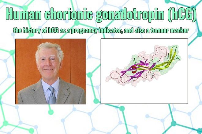
The history of human chorionic gonadotropin

Urine hCG as a pregnancy test
In the early years of the last century, the isolation of adrenalin and secretin and the concept of hormones as chemical messengers, as proposed by the British physiologist Ernest Starling in 1905, encouraged a surge of active endocrine research (most notably in Germany during the next three decades). This led to an improved understanding of the human ovarian cycle and the hormonal changes that take place in pregnancy.
In 1903, Ludwig Fraenkel, professor of gynaecology at the Women’s Hospital, Breslau provided experimental proof of the endocrine function of the corpus luteum. With the ready availability of placental tissue, studies by Bernhard Aschner (1912) and Otto Fellner (1913) and Japanese scientist Toyoichi Hirose (1920) showed placental tissue extracts had stimulatory effects on the genital tract, ovulation, corpus luteum and progesterone production of guinea pigs and rabbits (1). This demonstrated that the placenta had an endocrine function supporting pregnancy.
In 1928, German endocrinologist Selmar Aschheim and German-born Israeli gynaecologist Bernhard Zondek working at the Berlin Charite showed that pregnant women produced a high concentration of a gonad-stimulating substance (hCG) in their urine and that this activated receptors on the gonads of mice (2). Although the A-Z test took five days, was expensive and required special animal house facilities, it had a reported error rate of less than 2% and was sensitive in detecting hCG 7 to 10 days after a missed period.
A reference station was established in Edinburgh with a postal sample service in 1930 and the A-Z Test was soon used in some London hospitals. In 1931 Maurice Friedman and Maxwell Lapham at the University of Pennsylvania developed a similar technique with large female adult rabbits. The condition of the ovaries was examined after just one day so, by 1935, this had become the method of choice in London and the United States (3, 4).
hCG as a tumour marker
hCG is a 36.7kDa glycoprotein produced by the trophoblast tissue of the placenta in pregnancy and at a number of other sites in malignant conditions. It stimulates the ovarian corpus luteum to secrete progesterone until the placenta takes over production to sustain pregnancy. It also promotes cell fusion of cytotrophoblast to syncytiotrophoblast and maternal myometrial spiral artery angiogenesis.
hCG is a heterodimer with two glycosylated sub units; the alpha sub unit structure is common to other gonadotropins, for example luteinising hormone, but the beta sub unit of 145 amino acids is unique to hCG. The amino acid sequence of both sub units was established in 1975 by Francis Morgan and colleagues at Columbia University, New York.
In 1929, Aschheim (using the A-Z bioassay) reported greatly elevated urine hCG results in two forms of gestational trophoblastic disease (GTD), choriocarcinoma and hydatidiform mole, and in these cases urines often required 1/200 dilution (5). Choriocarcinoma was first described in Germany by Hans Chiari in 1877 with histological studies reported by M Saengers (1889) and six years later by Felix Marchand (6). It appears hydatidiform mole was recognised in ancient medicine but described in more detail by Madame Boivin in 1827, who recognised that the hydatids are grape-like cystic dilatations of the chorionic villi (7). Tumours of trophoblastic origin may arise in the uterus, placenta or gonads and tend to be highly malignant and may spread to the lungs and brain. Gestational choriocarcinoma (GC) is rare and occurs in 1/20-50,000 pregnancies, according to conflicting studies, but prior to the 1950s treatment was limited to surgery and radiation and the prognosis was poor. GC has a special place in cancer history, as it was the first condition successfully treated by chemotherapy when in 1958 Roy Hertz and Min Chiu Li at the National Institute of Health, Maryland developed the use of the folic acid inhibitor, methotrexate with great success (8, 9).
Clinical applications of hCG as tumour marker
The most often adult quoted reference range for serum hCG is
Serum hCG in combination with AFP and Lactate dehydrogenase is recognised as the best validated prognostic markers for diagnosis, follow up and monitoring germ cell tumours (seminomas, NSGCT and extragonadal tumours).
It is important to distinguish seminomas from teratomas, as each is treated differently with chemotherapy or surgery respectively. In seminomas hCG may range from 10-2000 IU/L. In NSGCT hCG may range from 5-1000 IU/L and depends on the stage of the tumour.
CSF hCG is more increased in primary intracranial germ cell tumours compared with serum hCG (10, 11).
Analysis of hCG
hCG exists in multiple forms in serum; intact hCG, Free Beta subunit, Beta core fragment, nicked hCG and hyperglycosylated hCG. Consequently, when used as a tumour marker the hCG assay should detect all main forms and laboratories should be aware of the characteristics of the assay in use in clinical interpretative medicine. The Beta core fragment is the main form in urine but hyperglycosylated hCG is the main urine form in early pregnancy.
The A-Z urine hCG test was used extensively with a number of modifications, most notably in 1941 by Eleanor Delfs, the distinguished professor of Obstetrics at Johns Hopkins University and later pioneer hCG researcher at Wisconsin Medical College. Delfs developed a serum hCG bioassay injecting serum extracts into female rats and measuring uterine weight (6, 12).
Bioassays continued until the first immunoassays were developed in 1960. This followed the typical development pattern of peptide immunoassays with first complement fixation. In the same year, a haemagglutination inhibition assay for urine hCG was devised by Lief Wide and Carl Gemzell in Stockholm. The reactants were incubated in tubes for two hours and read visually (13). In 1962, a group led by Jennifer Robbins developed a similar latex agglutination method using coated latex beads for urine hCG. The Gravindex pregnancy test was based on this latex agglutination inhibition principle.
In 1965, a radioimmunoassay was developed by a team at Charing Cross Hospital Medical School, led by CE Wilde (14). Charing Cross became a major oncology centre for GTD under the leadership of Prof Kenneth Bagshawe during this period and in 1969 he reported an improved hCG radioimmunoassay. Charing Cross was also notable for pioneering chemotherapy agent developments, including multidrug combinations under the leadership of Edward Newlands, in GTD, testicular, ovarian cancer and brain tumours.
Early commercial kit methods for urine hCG using the agglutination methods were compared and critically reviewed by Derek Watson in 1966 and found to be suitable for detection and monitoring GTD (15). In 1972, a radioimmunoassay specific for beta hCG was developed and was shown to be clinically useful in monitoring chemotherapy in GTD and follow up of terminated molar pregnancies (16). The introduction of monoclonal antibodies into immunoassays from 1975 led to a range of two antibody immunometric assays using high sensitivity fluorimetric and chemiluminescent tracer detection assays developed over the last few decades (11). Reported potential sources of error include lack of specificity. Some trophoblastic tumours secrete nicked hCG which is not measured in all assays. Due to the very high hCG concentrations often encountered the “high dose hook” effect produces false low results and samples require dilution. In addition, the presence of heterophilic antibodies may cause errors (10). Point-of-care and commercial devices for urine pregnancy testing generally are based on a sandwich ELISA dye detection principle and are qualitative only. It is essential that they detect hyperglycosylated hCG.
References
1. Hirose T: Exogenous stimulation of corpus luteum formation in the rabbit: influence of extracts of human placenta, decidua, fetus, hydatid mole, and corpus luteum on the rabbit gonad. J Jpn Gynecol Soc 1920, 16:1055
2. Aschheim S, Zondek B--The Diagnosis of Pregnancy From the Urine by Demonstration of the Hormone of the Pituitary Body. Klinische Wochenschrift, 7: 1404-1411, July 22, 1928
3. Olszynko-Gryn J. The demand for pregnancy testing. Stud Hist Philos Biol Biomed Sci 2014; 47: 233-247
4. Friedman MH, Lapham M. A simple, rapid procedure for the diagnosis of early pregnancies. Am J Obs Gynae 1931; 21: 405-410
5. Aschheim S. The early diagnosis of pregnancy, chorio-epithelioma and hydatidiform mole by the Aschheim-Zondek test. Amer J Obs Gynae. 1929; 28: 335-342
6. Cole L, Butler S. Human chorionic gonadotrophin, 2nd Edition, 2015 published by Elsevier, London
7. Hunter J. Treatment of hydatidiform mole. Surg Clin North America 1959; 39(4): 1131-1136
8. Chabner BA, Roberts TG Jr. Perspectives-chemotherapy and the war on cancer. Nature Reviews Cancer. 2005; 5
9. Li MC, Hertz R, Bergenstal DM. Therapy of choriocarcinoma and related trophoblastic tumours with folic acid and purine antagonists. New Engl J Med 1958; 259: 66-74
10. Human Chorionic Gonadotrophin. Association for Clinical Biochemistry and Laboratory Medicine 2014.
11. Cole L New discoveries on the biology and detection of human chorionic gonadotrophin Repro Biol End 2009; 7(1):8
12. Delfs E. An assay method for Human Chorionic Gonadotrophin. Endocrin 1941; 28: 196-202
13. Wide L, Gemzell CA. An immunological pregnancy test. Acta Endocrinologica 1960; 35: 261-267
14. Wilde CE, Orrs AH, BagshaWe KD. A radioimmunoassay for human chorionic gonadotrophin. Nature 1965; 205: 191-192
15. Watson D. Urinary human chorionic gonadotrophin. Clan Chem 1966; 12: 577-585
16. Vaitukaitis J, Braunstein GD, Ross GT. A radioimmunoassay which specifically measures human chorionic gonadotrophin in the presence of human luteinising hormone. Am J Obs & Gynae 1972; 113(6): 751-758
