Biomedical scientists at the forefront of cancer detection
The number of diagnosed cancer cases is growing. Over 360,000 people in the UK have been diagnosed with cancer within the last two years, at an average of more than 800 new cases a day.
Early diagnosis is essential for treating cancer. Cancer survival rates are improving and have doubled in the last forty years, with half of patients diagnosed with the 12 most common cancers surviving for ten years or more.
A large number of cancer diagnoses are made or confirmed using a tissue biopsy, which is then sent to a pathology laboratory for analysis and reporting. Laboratories operate under strict rules regarding the handling and storage of human tissue.
The Human Tissue Act of 2004 was introduced to address ethical issues relating to the legal removal, storage, use and disposal of human tissues, organs and bodies. The Human Tissue Authority (HTA), is the regulator that ensures that clinical standards are set and adhered to, whereby a tissue sample is only taken and handled with a patent’s consent.
Tissue samples can only be used for research purposes if there is patient consent, or where the tissue is released to the researcher in a non-identifiable form to be used in a Research Ethics Committee approved project.
It is important to appreciate that taking even the smallest biopsies is an invasive procedure and carries a degree of risk, albeit usually a very small one. An exciting piece of research involving biomedical scientists is the development of alternative techniques to tissue sampling for diagnosis that uses biophotonics and Raman spectroscopy.
Alternative methods of tissue analysis: biophotonics and Raman spectroscopy
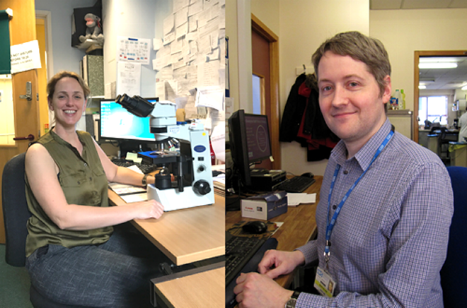
Joanne Motte is a senior biomedical scientist and an advanced practitioner in histological dissection at Gloucestershire Hospitals NHS Foundation Trust. Joanne works with the hospital’s Biophotonics Research Unit; a team of clinicians, clinical scientists, information technologists and biomedical scientists who implement photon based technological methods for tissue analysis.
Her team is looking at alternative approaches to the conventional tissue biopsy for sites that are deep within the body and inaccessible. Joanne explains:
“A biopsy is not appropriate if the disease is in a vulnerable area and cannot be sampled. Obtaining a tissue sample for diagnosis could cause harm to the patient and potentially serious complications after the specimens are removed, and there are areas in the body where it is simply too difficult and risky to attempt a biopsy.
The Biophotonics Unit is researching novel methods of disease detection; these are near the patient tests and provide information immediately, allowing directed treatment without destroying or removing any tissue as a biopsy.”
One method of analysing suspected cancerous tissue is Raman spectroscopy, a technique using a spectrum of scattered light to produce a chemical fingerprint of tissue.
Joanne added, “Raman spectroscopy can potentially be used to obtain an almost instant diagnosis of cancers and pre-cancers in vivo by detection of the biochemical markers.”
Once cancerous tissue has been identified, Raman spectroscopy can distinguish between early malignancies and benign growths, and detect abnormalities in specific areas of tissue which can then be treated.
Joanne’s team is working to establish the possibility of performing these analyses, which would lead to an ‘optical biopsy’, providing information on cancerous cells without removing the tissue from the patient.
Joanne said, “This would preclude the need for a biopsy, making it potentially a one stop clinic format. This technique has shown diagnostic potential in the oesophagus and also in the measuring and diagnosis of malignant pathologies in lymphatic systems, breast, prostate and vulval skin tissue. The concept is to see and treat in one visit.”
To confirm the accuracy of Raman spectroscopy in identifying abnormal cells and tissues, Joanne uses conventional microscopic assessment of tissues in parallel to compare with the spectroscopy results for consistency.
She concluded, “The application of this exciting science holds enormous potential for improving the diagnosis and treatment of disease, including cancer.”
Whilst these methods are exciting advancements in the field of diagnostic research, other methods like digital pathology require biopsies but offer equal potential for improving cancer diagnostic services.
Digital pathology and artificial intelligence
Ashley Ballard is a senior specialist biomedical scientist in the histopathology department at The Royal Bournemouth Hospital. He has previously published research on breast cancer and is looking at how digital pathology can facilitate cancer diagnostic services for his PhD.
Digital pathology uses digitally scanned images of standard glass slides produced in the histology laboratory. These whole slide images allow expert opinions to be sought instantly on any challenging case, reducing turnaround times.
Another potentially transformative benefit to digital pathology is that it allows the use of image analysis, which is a form of artificial intelligence.
Ashley said:
“By using supervised learning on deep neural networks, computers can be trained to recognise certain tissue features, allowing cell counting and other repetitive and time-consuming tasks currently carried out by pathologists to be automated.
At present, digital pathology is only used for primary diagnosis in a small number of UK laboratories. However, as the usage of these techniques spreads and new image analysis protocols become available, it could have a significant effect on the speed of diagnosis, repeatability of testing, and the quality of diagnostic reports. The main focus of my research is to quantify and model these benefits, to determine if smarter, better, cheaper, faster, and more reliable cancer diagnostic services can be made available through the use of digital pathology.
The majority of research in this area uses supervised learning to focus on the development of computer aided diagnostic tools to aid the pathologist. These will allow the quantification of the extent and intensity of a range of other markers, providing more information to inform the diagnosis as well as aiding the detection of features that might otherwise have been missed.
Although this will provide tangible benefits to cancer detection and diagnosis, one of my longer-term research goals is to investigate the viability of semi-automated diagnosis, as well as the use of un-supervised machine learning to develop entirely computer generated diagnostic categories.
This will require harnessing big data (scanning hundreds of thousands of slides) as well as existing multiplex staining techniques, providing insights human pathologists using standard techniques would not normally be able to perceive. This will add additional layers of diagnostic detail to that provided by the pathologist, such as information on the tumour micro-environment, thus improving cancer outcomes.”
With these new and emerging technologies coming into play in the laboratory, biomedical scientists are making advances in cancer diagnostic services, however more research is needed.
World Cancer Day
4th February marks World Cancer Day, following the theme of ‘We can. I can.’ This year charities and professional bodies like Cancer Research UK are asking people to take part in their World Cancer Day campaign and to show their support of cancer research.
Macmillan Cancer Support estimates that by 2020, almost one in two people will develop cancer during their lifetimes.
Help raise awareness and show your support for cancer research today. You can follow the campaign on Twitter using the hashtags #WorldCancerDay and #WeCanICan.