Inside the lab: childhood cancer treatment
Being diagnosed with cancer can be frightening but when the patient receiving the diagnosis is a child, it can be heart-breaking.
It is estimated that more than 1400 new cases of childhood cancer are diagnosed each year in the UK. The biggest cancers affecting children are leukaemia, lymphomas, sarcomas and nervous cell tumours, with children most at risk of developing cancer in the first five years of life.
A 2018 Public Health England report states that, of all diseases, cancer is the most common cause of death in children aged one to fourteen years old, accounting for a fifth of all deaths in this age bracket. Whilst there is a high survival rate for children’s cancers (82% of diagnosed child patients survive their disease for five years or more), due to their rarity these diseases are often misunderstood.
When a child is first suspected to have cancer, their blood, bone marrow or tissue sample is examined by biomedical scientists in a laboratory. Through a series of analysis like immunophenotyping by flow cytometry, histology and molecular screening, scientists determine whether the sample is cancerous and which type of cancer it is. This enables the clinician to begin the correct treatment protocol specific to the cancer type.
To learn more about the processes behind diagnosing and treating paediatric cancer patients, we spoke with three biomedical scientists at Great Ormond Street Hospital (GOSH).
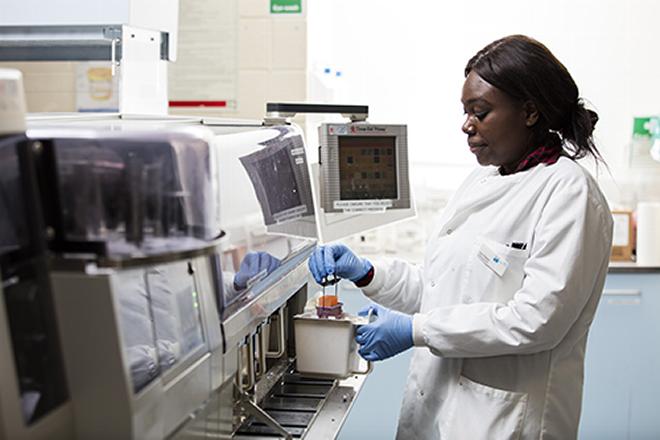
Diagnosing cancerous cells through – Immunophenotyping using flow cytometry
GOSH has a lab dedicated to haematological and solid tumour immunophenotyping by flow cytometry. This laboratory is part of the Specialist Integrated Haematological Malignancy Diagnostic Services (SHIHMDS) department. The scientists in this laboratory study cells from various sample types using a cell labelling process known as immunophenotyping.
Lead Healthcare Scientist Sarah Inglott said, “The biomedical scientists in the lab are responsible for setting up the analysis, acquiring the data, analysing and interpreting the results, completing a report and authorising the result. Flow cytometry plays an integral role in diagnostic pathology and monitoring of treatment responses during patient therapies.”
Immunophenotyping labels cell antigens found on the surface or inside the cell. This process can be used for many cells types. For instance, a normal white cell will express these antigens at a specific level throughout its maturation. In malignancies such as leukaemia the antigen will be expressed at abnormal levels (or not in synchrony) and this is known as an aberrant phenotype.
Sarah explained, “At diagnosis when abnormal cells are identified, an aberrant phenotypic pattern is created for each patient. These abnormal phenotypes can be tracked throughout treatment to ascertain disease clearance and measure how well a patient is responding to treatment. They can also be used in long term follow up to detect early relapse before the patient becomes clinically unwell. This allows for either treatment intensification, choice to go ahead with transplant or other immunotherapy options.
Specialised treatments for child cancer patients
In the Molecular Minimal Residual Disease laboratory (MRD), biomedical scientists monitor disease progress and the effectiveness of chemotherapy in patients with acute lymphoblastic leukaemias (ALL).
Specialist Translational Biomedical Scientist Eleanor Watt said, “Molecular MRD testing involves bespoke assays to be designed for each and every patient under our care. This involves a huge amount of work for the biomedical scientists working in this department.
The biomedical scientists examine the different re-arrangements of DNA in cancer cells affecting a patient’s white blood cells. When a new diagnostic sample arrives, we have to screen the patient, which involves extracting and copying the DNA, running the products on a gel, cutting out the bands that appear for the patient, cleaning them up and purifying them.”
An experienced biomedical scientist then examines the sequenced products, recognises where the clonal rearrangement(s) are and design primers, which are precise DNA sequences that will only attach to the rearrangement in this patient. This means that every child with ALL has their own unique assay developed.
Eleanor concludes, “Samples from the child are sent at regular time points, to ensure the treatment is working, meaning the doctors rely heavily on the biomedical scientists for accurate results in order to treat the patient appropriately.”
Monitoring patients’ treatment
Treatments used for fighting cancers include chemotherapy, surgery, radiotherapy, precision medicine and more. Whilst there are many therapies and treatments available, the first line of treatment might not work, causing some patients to require a bone marrow or blood stem cell transplant (BMT).
Inside the SIHMDS department at GOSH is the molecular haematology laboratory, where the biomedical scientists monitor patients who have had bone marrow transplants.
Senior Specialist Biomedical Scientist Susanne Kricke commented, “To see if the BMT has been successful, biomedical scientists monitor the patient’s engraftment. To do so, the genetic fingerprints of the patient and the donor are established prior to the BMT. This is done by testing their DNA and looking for differences in repetitive sequences within the DNA. The two original genetic fingerprints from the patient and the donor are then compared to the genetic fingerprint of the patient’s blood or bone marrow after the transplant.
This way, the biomedical scientist can tell how many patient cells and how many donor cells are in the sample and if the BMT has worked or not. The engraftment is regularly monitored by the biomedical scientists and any changes in the patient’s engraftment are communicated to the doctors urgently so that treatment can be adjusted accordingly.”
During Childhood Cancer Awareness Month, we want to thank our members for the vital role they play at the heart of every child’s healthcare: from their in-depth analysis of the samples in the laboratory, and identifying and diagnosing cancerous samples, to designing personalised treatments and monitoring patients’ reactions to treatment on a cellular and DNA level.